Meningioma: Radiation Therapy Overview
English Version
1. Indications for Radiation Therapy
Meningiomas are typically benign, slow-growing tumors arising from the meninges, the membranes surrounding the brain and spinal cord. Radiation therapy is considered in several scenarios:
- Post-Surgical Adjuvant Therapy: Following subtotal resection where residual tumor remains, especially if complete resection is not possible due to tumor location.
- Recurrent Meningioma: When the tumor recurs after surgery.
- Unresectable Tumors: Primary treatment for tumors that cannot be surgically removed due to their location or the patient's condition.
- Atypical or Malignant Meningioma: High-grade meningiomas (WHO Grade II or III) are more aggressive, and adjuvant radiation is often recommended even after gross total resection.
2. Treatment Field
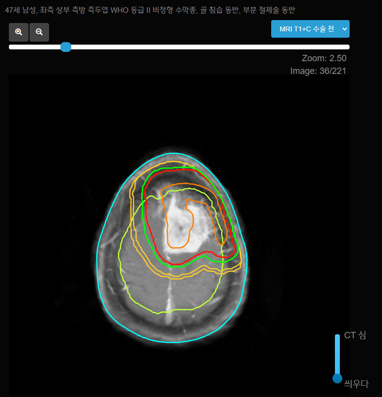
- Gross Tumor Volume (GTV): Includes the visible tumor on MRI and any residual tumor after surgery.
- Clinical Target Volume (CTV): A margin of 0.5-1 cm is typically added to the GTV to account for microscopic disease extension.
- Planning Target Volume (PTV): A further margin of 0.3-0.5 cm is added to the CTV to account for setup uncertainties and patient movement during treatment.
3. Radiation Dose
- Benign Meningioma (WHO Grade I): Standard dose ranges from 50.4 Gy to 54 Gy, delivered in fractions of 1.8 Gy over 6 weeks.
- Atypical Meningioma (WHO Grade II): A higher dose of 54-60 Gy is typically delivered in fractions of 1.8-2 Gy.
- Malignant Meningioma (WHO Grade III): These aggressive tumors may require doses up to 60-66 Gy, also delivered in fractions of 1.8-2 Gy.
- Stereotactic Radiosurgery (SRS): For small meningiomas, particularly those less than 3 cm in size or in critical locations, a single high-dose fraction (12-16 Gy) or hypofractionated radiosurgery (e.g., 25 Gy in 5 fractions) may be used.
4. Treatment Techniques
- 3D Conformal Radiation Therapy (3D-CRT): Often used to precisely target the tumor while sparing surrounding healthy tissue.
- Intensity-Modulated Radiation Therapy (IMRT): Offers superior dose distribution, particularly for tumors near critical structures like the optic nerves or brainstem.
- Stereotactic Radiosurgery (SRS): Highly focused radiation used for small or well-circumscribed tumors, allowing for the delivery of high doses to the tumor while minimizing exposure to surrounding tissue.
5. Side Effects
- Acute Effects: Fatigue, mild skin reactions at the treatment site, temporary hair loss, and headaches.
- Late Effects: Depending on the tumor location, late effects may include cognitive decline, radionecrosis, or cranial nerve deficits, although these are less common with precise targeting techniques like IMRT or SRS.
6. Follow-up
- Imaging: Regular MRI scans post-treatment to monitor for tumor control or recurrence.
- Clinical Assessment: Ongoing evaluation of neurological function, especially in cases where the tumor is near critical structures such as the optic nerves or cranial nerves.
Korean Version
1. 방사선치료 적응증
수막종은 일반적으로 양성이고, 천천히 성장하는 종양으로 뇌와 척수를 둘러싼 막(수막)에서 발생합니다. 방사선 치료는 다음과 같은 경우에 고려됩니다:
- 수술 후 보조 치료: 종양이 불완전하게 절제된 경우, 특히 종양의 위치로 인해 완전 절제가 불가능한 경우.
- 재발성 수막종: 수술 후 종양이 재발한 경우.
- 수술 불가능한 종양: 종양의 위치나 환자의 상태로 인해 수술이 불가능한 경우의 주요 치료법.
- 비정형 또는 악성 수막종: 보다 공격적인 고등급 수막종(WHO 2급 또는 3급)에서는 종양이 완전 절제된 후에도 보조 방사선치료가 권장됩니다.
2. 치료 범위
- 종양 부피(GTV): MRI에서 확인된 종양과 수술 후 남아 있는 종양을 포함합니다.
- 임상 표적 부위(CTV): 미세 질병 확장을 고려하여 GTV에 보통 0.5-1cm의 마진을 추가합니다.
- 계획 표적 부위(PTV): 치료 중 설정 불확실성과 환자의 이동을 보완하기 위해 CTV에 추가적으로 0.3-0.5cm의 여유 마진을 더한 것입니다.
3. 방사선 선량
- 양성 수막종(WHO 1급): 표준 선량은 50.4 Gy에서 54 Gy 사이이며, 1.8 Gy씩 6주 동안 투여됩니다.
- 비정형 수막종(WHO 2급): 일반적으로 54-60 Gy의 높은 선량을 1.8-2 Gy씩 분할하여 투여합니다.
- 악성 수막종(WHO 3급): 이러한 공격적인 종양은 60-66 Gy의 선량이 필요할 수 있으며, 1.8-2 Gy씩 분할하여 투여합니다.
- 정위 방사선 수술(SRS): 크기가 3cm 이하이거나 중요한 위치에 있는 작은 수막종의 경우, 단일 고용량 분할(12-16 Gy) 또는 저분할 방사선 수술(예: 25 Gy/5회)로 치료할 수 있습니다.
4. 치료 기법
- 3차원 적형 방사선치료(3D-CRT): 종양을 정밀하게 타겟팅하면서 주변 건강한 조직을 보호할 수 있는 방법으로 자주 사용됩니다.
- 강도 변조 방사선치료(IMRT): 특히 시신경이나 뇌간과 같은 중요한 구조물 근처의 종양에서 우수한 선량 분포를 제공합니다.
- 정위 방사선 수술(SRS): 작고 잘 구획된 종양에 사용되는 고집중 방사선으로, 종양에 높은 선량을 전달하면서 주변 조직에 대한 노출을 최소화합니다.
5. 부작용
- 급성 부작용: 피로, 치료 부위에서의 가벼운 피부 반응, 일시적인 탈모, 두통 등이 있습니다.
- 지연된 부작용: 종양 위치에 따라 지연된 부작용으로 인지 기능 저하, 방사선 괴사, 뇌신경 결손 등이 있을 수 있지만, IMRT 또는 SRS와 같은 정밀 타겟팅 기법을 사용하면 이러한 위험은 줄어듭니다.
6. 추적 관리
- 영상 검사: 치료 후 정기적으로 MRI 스캔을 통해 종양 조절 또는 재발 여부를 모니터링합니다.
- 임상 평가: 특히 시신경 또는 뇌신경과 같은 중요한 구조물 근처에 있는 경우 신경학적 기능의 지속적인 평가가 필요합니다.


Meningioma: Radiation Therapy Overview
English Version
1. Indications for Radiation Therapy Meningiomas are typically benign, slow-growing tumors arising from the meninges, the membranes surrounding the brain and spinal cord. Radiation therapy is considered in several scenarios:
2. Treatment Field
3. Radiation Dose
4. Treatment Techniques
5. Side Effects
6. Follow-up
Korean Version
1. 방사선치료 적응증 수막종은 일반적으로 양성이고, 천천히 성장하는 종양으로 뇌와 척수를 둘러싼 막(수막)에서 발생합니다. 방사선 치료는 다음과 같은 경우에 고려됩니다:
2. 치료 범위
3. 방사선 선량
4. 치료 기법
5. 부작용
6. 추적 관리