Pituitary Adenoma: Radiation Therapy Overview
English Version
1. Indications for Radiation Therapy Pituitary adenomas are generally benign tumors of the pituitary gland, but they can cause significant clinical symptoms due to hormone overproduction or mass effect on surrounding structures such as the optic chiasm. Radiation therapy is considered in the following scenarios:
- Post-Surgical Adjuvant Therapy: After incomplete resection, especially if the tumor is invasive or near critical structures like the optic nerves.
- Recurrent or Residual Tumor: When there is evidence of tumor recurrence or residual tumor that is not amenable to further surgery.
- Non-Functioning Pituitary Adenomas: For non-functioning adenomas causing mass effect that are not completely resectable.
- Hormone-Secreting Adenomas: When medical therapy (e.g., dopamine agonists for prolactinomas) and surgery fail to control hormone hypersecretion.
2. Treatment Field
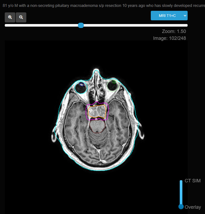
- Gross Tumor Volume (GTV): Includes the visible tumor on MRI and any residual tumor post-surgery.
- Clinical Target Volume (CTV): Typically includes a margin of 2-3 mm around the GTV to account for microscopic disease spread.
- Planning Target Volume (PTV): An additional 1-2 mm margin is added to the CTV to accommodate patient movement and setup uncertainties.
3. Radiation Dose
- Conventional Fractionation: For most pituitary adenomas, a dose of 45-54 Gy delivered in 1.8-2 Gy fractions over 5-6 weeks is standard.
- Stereotactic Radiosurgery (SRS): A single high-dose fraction (12-20 Gy) is used for smaller tumors (<3 cm) or those in critical locations, offering a highly targeted approach that minimizes exposure to surrounding tissues.
- Hypofractionated Stereotactic Radiotherapy (HSRT): For tumors that are larger or adjacent to critical structures, a hypofractionated regimen (e.g., 25 Gy in 5 fractions) may be employed.
4. Treatment Techniques
- 3D Conformal Radiation Therapy (3D-CRT): Commonly used for its ability to conform the radiation dose to the shape of the tumor while sparing surrounding healthy tissue.
- Intensity-Modulated Radiation Therapy (IMRT): Provides superior dose distribution, particularly beneficial for tumors near critical structures like the optic nerves and brainstem.
- Stereotactic Radiosurgery (SRS): Ideal for small, well-defined adenomas, delivering a high dose to the tumor with minimal exposure to nearby critical structures.
- Proton Therapy: Considered for select cases where reducing radiation dose to surrounding tissues, particularly in younger patients, is a priority.
5. Side Effects
- Acute Effects: Fatigue, mild headaches, and transient nausea. Localized skin reactions and hair loss may occur if the treatment field includes skin surfaces.
- Late Effects: Potential long-term effects include hypopituitarism (requiring lifelong hormone replacement therapy), optic neuropathy, or very rarely, secondary malignancies. These risks are generally lower with advanced techniques like IMRT or SRS.
6. Follow-up
- Imaging: Regular MRI scans post-treatment to monitor for tumor control or recurrence.
- Endocrine Function Testing: Periodic assessment of pituitary hormone levels to detect and manage hypopituitarism or other endocrine dysfunctions.
- Ophthalmologic Evaluation: Regular eye exams to monitor for any vision changes, especially if the tumor was near the optic chiasm.
Korean Version
1. 방사선치료 적응증
뇌하수체 선종은 일반적으로 양성 종양이지만, 호르몬 과다 분비 또는 시신경교차와 같은 주변 구조물에 대한 압박으로 인해 중요한 임상 증상을 유발할 수 있습니다. 방사선 치료는 다음과 같은 경우에 고려됩니다:
- 수술 후 보조 치료: 종양이 불완전하게 절제된 후, 특히 종양이 침습적이거나 시신경과 같은 중요한 구조물 근처에 있는 경우.
- 재발성 또는 잔류 종양: 종양이 재발하거나 추가 수술이 어려운 잔류 종양이 있는 경우.
- 비기능성 뇌하수체 선종: 완전 절제가 불가능한 압박 효과를 유발하는 비기능성 선종.
- 호르몬 분비 선종: 의료 요법(예: 프로락틴종의 경우 도파민 작용제)과 수술이 호르몬 과다 분비를 조절하지 못하는 경우.
2. 치료 범위
- 종양 부피(GTV): MRI에서 보이는 종양과 수술 후 남아 있는 종양을 포함합니다.
- 임상 표적 부위(CTV): GTV 주위에 미세 질병 확산을 고려하여 2-3 mm의 마진을 추가합니다.
- 계획 표적 부위(PTV): 환자 이동과 설정 불확실성을 보완하기 위해 CTV에 추가적으로 1-2 mm의 여유 마진을 더한 것입니다.
3. 방사선 선량
- 표준 분할: 대부분의 뇌하수체 선종의 경우, 45-54 Gy의 선량을 1.8-2 Gy씩 5-6주 동안 투여하는 것이 표준입니다.
- 정위 방사선 수술(SRS): 작은 종양(<3 cm) 또는 중요한 위치에 있는 종양의 경우, 단일 고용량 분할(12-20 Gy)로 치료하며, 이는 주변 조직에 대한 노출을 최소화하면서 종양에 높은 선량을 전달하는 고도로 표적화된 접근법입니다.
- 저분할 정위 방사선치료(HSRT): 더 큰 종양이나 중요한 구조물에 인접한 종양의 경우, 저분할 일정(예: 25 Gy/5회)을 사용할 수 있습니다.
4. 치료 기법
- 3차원 적형 방사선치료(3D-CRT): 종양의 형태에 맞추어 방사선 선량을 조절하면서 주변 건강한 조직을 보호할 수 있어 자주 사용됩니다.
- 강도 변조 방사선치료(IMRT): 시신경 및 뇌간과 같은 중요한 구조물 근처의 종양에 특히 유용한 우수한 선량 분포를 제공합니다.
- 정위 방사선 수술(SRS): 작고 잘 정의된 선종에 이상적인 치료법으로, 주변의 중요한 구조물에 대한 노출을 최소화하면서 종양에 높은 선량을 전달합니다.
- 양성자 치료: 특히 젊은 환자에게 주변 조직에 대한 방사선 선량을 줄이기 위해 선택적으로 고려될 수 있습니다.
5. 부작용
- 급성 부작용: 피로, 가벼운 두통, 일시적인 메스꺼움 등이 있으며, 치료 범위에 피부가 포함된 경우 국소적인 피부 반응 및 탈모가 발생할 수 있습니다.
- 지연된 부작용: 장기적인 효과로는 뇌하수체 기능 저하(평생 호르몬 대체 요법이 필요할 수 있음), 시신경병증, 드물게 이차 암 발생 등이 있을 수 있습니다. 이러한 위험은 IMRT 또는 SRS와 같은 고급 기술을 사용하면 일반적으로 낮습니다.
6. 추적 관리
- 영상 검사: 치료 후 정기적으로 MRI 스캔을 통해 종양 조절 또는 재발 여부를 모니터링합니다.
- 내분비 기능 검사: 뇌하수체 기능 저하 또는 기타 내분비 기능 장애를 발견하고 관리하기 위해 주기적인 뇌하수체 호르몬 수치 평가가 필요합니다.
- 안과 검사: 특히 종양이 시신경교차 근처에 있는 경우 시력 변화를 모니터링하기 위해 정기적인 안과 검사가 필요합니다.


Pituitary Adenoma: Radiation Therapy Overview
English Version
1. Indications for Radiation Therapy Pituitary adenomas are generally benign tumors of the pituitary gland, but they can cause significant clinical symptoms due to hormone overproduction or mass effect on surrounding structures such as the optic chiasm. Radiation therapy is considered in the following scenarios:
2. Treatment Field
3. Radiation Dose
4. Treatment Techniques
5. Side Effects
6. Follow-up
Korean Version
1. 방사선치료 적응증 뇌하수체 선종은 일반적으로 양성 종양이지만, 호르몬 과다 분비 또는 시신경교차와 같은 주변 구조물에 대한 압박으로 인해 중요한 임상 증상을 유발할 수 있습니다. 방사선 치료는 다음과 같은 경우에 고려됩니다:
2. 치료 범위
3. 방사선 선량
4. 치료 기법
5. 부작용
6. 추적 관리